The xiphisternal joint is at the apex of the infrasternal angle, which is formed by costal cartilage 7 on each side.
What Is The Inferior Wall Of The Heart. Can it be detected with echocardiography? It arises from the lower part of the interventricular septum and crosses the interior space of the right ventricle to connect with the inferior papillary muscle.7 the right ventricle tapers into the pulmonary.
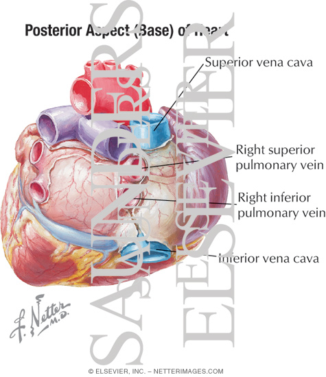
Posterior Aspect (base) of Heart With Light Micrograph of ... from www.netterimages.com
The heart is a muscular organ that pumps blood throughout the body. Part of the inside walls of the ventricles. Inside, the heart is divided into four heart chambers:
The human heart functions throughout a person's lifespan and is one of the most robust and hardest working muscle in the human body.
The superior vena cava and inferior vena cava drain systemic venous blood into the posterior wall of the right atrium. The pulmonary veins take blood from the lungs to the left atrium. The heart is located between the two lungs in the space referred to as the mediastinum (the space in the chest between the pleural sacs of the lungs that 3. Explore its structure, functions and facts only at byju's biology.